Read more at: Hope, Science, and Solidarity: A rare cancer community comes together
Hope, Science, and Solidarity: A rare cancer community comes together
9 July 2025
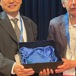
On Friday, 27th June 2025, a unique and powerful gathering took place in Cambridge. For the first time, mothers, researchers, and a young adult patient—who had long supported each other online—came together in person to focus on a shared mission: improving the understanding and treatment of anaplastic large cell lymphoma (...