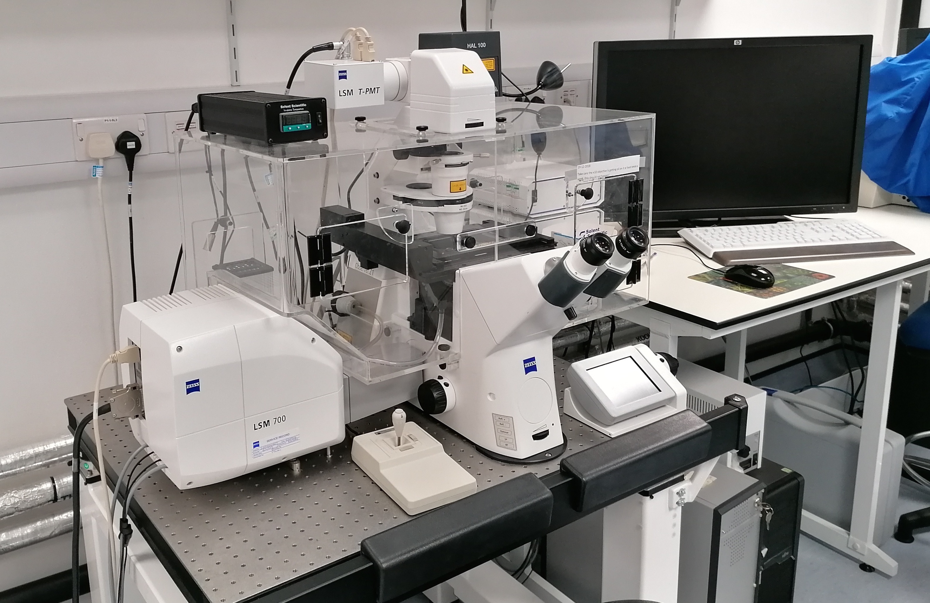
Inverted microscope compatible for 4-channel imaging and incubation chamber for live-cell imaging. Our incubation chamber allows control over temperature, CO2 and humidity to permit live-cell imaging experiments.
Features:
* 3D imaging
The LSM 700 assists you in configuring the acquisition parameters, from choosing the pixel resolution, via setting the diameter of the confocal pinhole to the Z spacing of the optical sections. Subsequently image acquisition is performed automatically and fully motorized. The ZEN software reconstructs your highly resolved 3D images and meaningfully presents them, e.g., in the form of projection or animations.
* Four-channel confocal microscopy
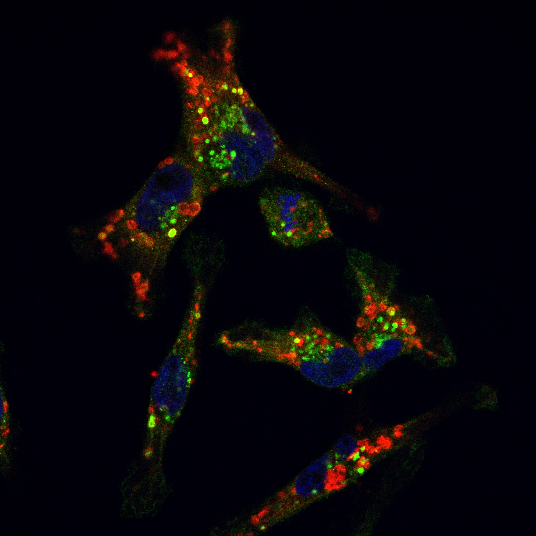
Multiple fluorescence and colocalization analyses In multicolor fluorescence imaging, the use of several fluorophores permits the observation of spatial relations between several cell constituents. 2 fluorescence detectors in the LSM 700 detect up to four color signals in a (quasi-)simultaneous mode, at frame rates of up to 5 fps for 512 x 512 pixels. Efficient separation of the fluorescence signals by selective laser excitation, and efficient splitting by means of the VSD (Variable Secondary Dichroic) beamsplitter prevent crosstalk and ensure unambiguous results, especially in colocalization analyses.
* Emission Fingerprinting
Spectral imaging and subsequent linear unmixing precisely separate fluorescent signals even of greatly overlapping color signals – whether you use, for example, GFP and YFP simultaneously or whether broad-band autofluorescence is present.
* Live Cell Imaging
High light intensities and long irradiation lead to phototoxic reactions in living cells and tissues. The high sensitivity of the LSM 700, combined with pixel-precise control of illumination, preserves your specimens and permits you to observe fast biological processes over long periods of time.
* FRAP, FLIP, photoactivation and photoconversion
Transport processes in live cells and organisms can be observed by means of targeted localized photobleaching, or by means of photoactivation or colour conversion of fluorophores such as PA-GFP or Kaede.
* Image analysis features on ZEN software including colocalisation, intensity measurements.
Objectives: 10x (air), 20x (air), 40x (air), 63x (oil), 100x (oil)
Scan Resolution: 2048 x 2048 pixels (max).
Software: ZEN black
(free downloads available from ZEN website for image analysis on your PC)


