Read more at: Are aesthetic exosome treatments dangerous?
Are aesthetic exosome treatments dangerous?
30 July 2025
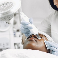
In recent years, exosome therapy has emerged as a hot trend in the world of aesthetic medicine. Marketed as a regenerative, cutting-edge solution for anti-ageing, skin rejuvenation, hair restoration, and even scar reduction, exosome treatments are often pitched as a safer, more effective alternative to traditional cosmetic...