
 A new study led by Dr Richard Hayward illuminating how the pathogenic bacterium Chlamydia trachomatis enters mammalian host cells has just been published in PLoS Pathogens.
C.trachomatis remains the leading bacterial agent of sexually transmitted disease worldwide and causes a form of blindness (trachoma) in Developing nations, which is recognised as a neglected tropical disease. Despite this burden, we know comparatively little about how it causes disease at a molecular level.
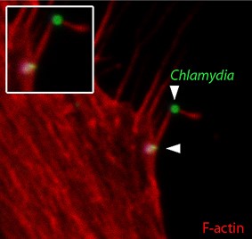
Building on a previous cryo-electron tomography study [Nans et al (2014) Cellular Microbiology 16:1457], this new work describes previously unrecognised events early during Chlamydia-host interaction, including the capture of extracellular bacteria by actin-rich protrusions called filopodia, and uncovered an induced-entry pathway sharing key hallmarks with cellular macropinocytosis. This revealed an unanticipated complexity of signalling underpinning cell entry by this major human pathogen, and suggests intriguing parallels with viral mechanisms of cell entry.
This new work is part of a wider project analysing Chlamydia-host interactions, and was funded by the Medical Research Council.
Chlamydia exploits filopodial capture and a macropincocytosis-like pathway for host cell entry
Ford C, Nans A, Boucrot E, Hayward RD
PLoS Pathogens 14:e1007051
http://journals.plos.org/plospathogens/article?id=10.1371/journal.ppat.1007051