Alt Text:
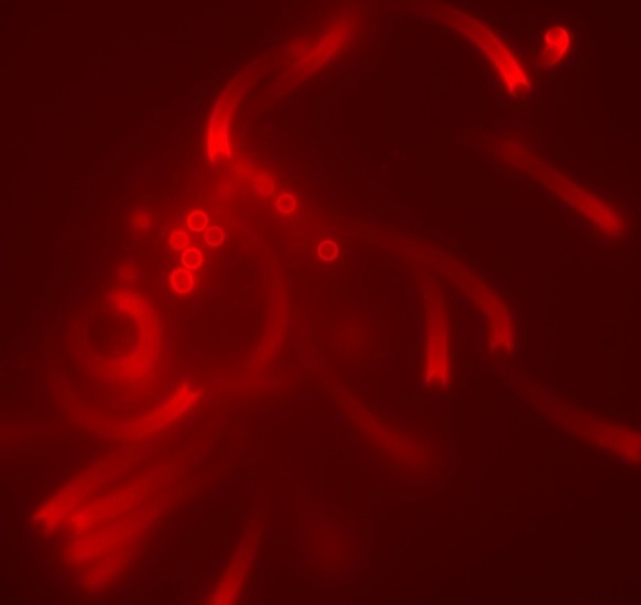
Figure: Phospholipid membrane-coated beads, incubated in a brain cell extract, assemble actin "comet tails" (red) which propel the beads through the extract.
Title Text:
Phospholipid membrane-coated beads

Department of Pathology

Department of Pathology
University of Cambridge
Tennis Court Road
Cambridge
CB2 1QP
+44 (0)1223 333690

© 2025 University of Cambridge